
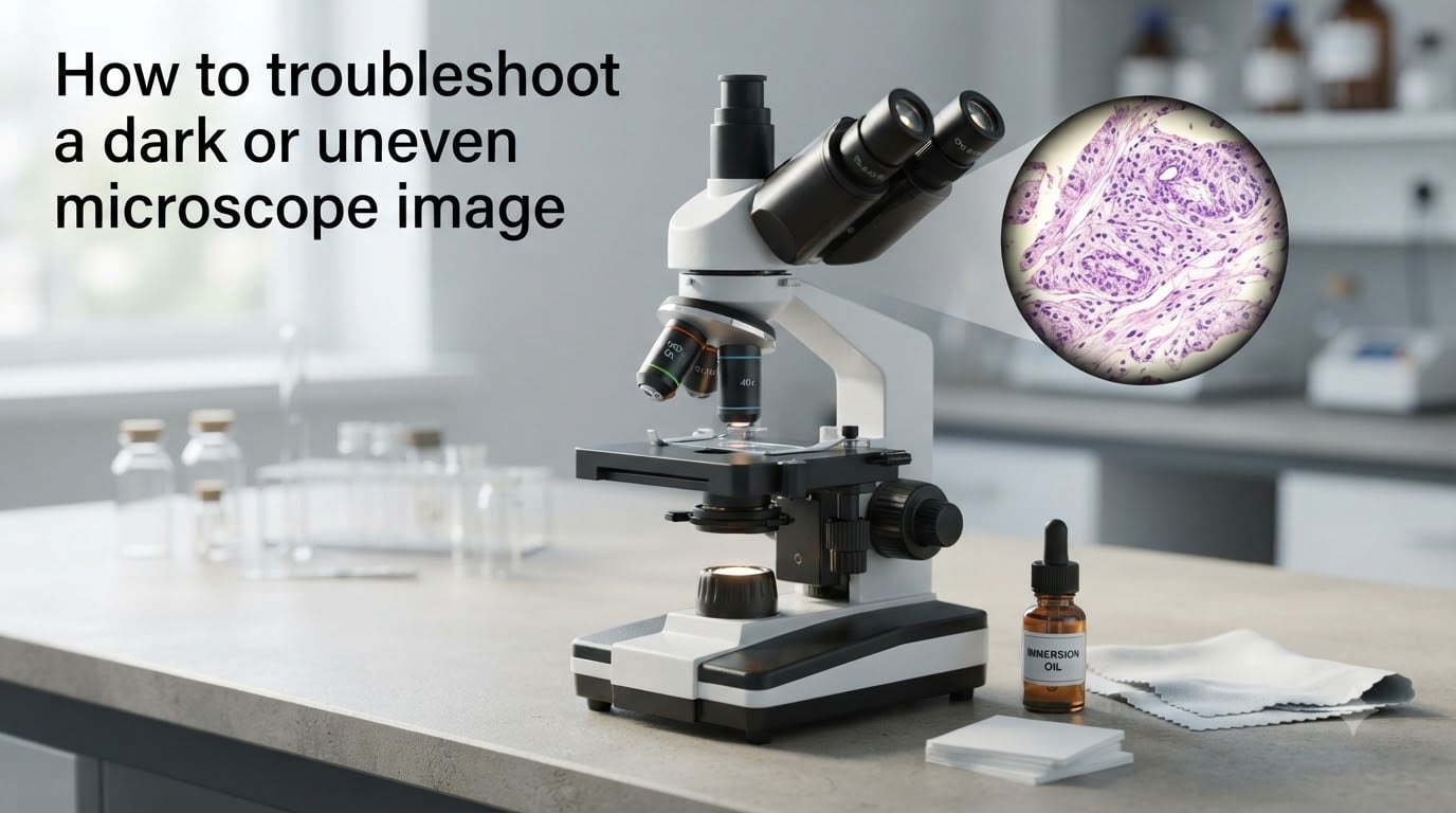
If your microscope image looks darker than usual, has dim corners, or shows a bright hotspot that you cannot seem to balance, you are not alone. I have watched new lab members lose entire afternoons chasing a single dirty objective or a lamp filament that drifted out of position. The good news is that almost every dark or uneven microscope image can be fixed by working through a short, repeatable checklist.
This guide shows you how to troubleshoot a dark or uneven microscope image in the order that solves most problems fastest. I will walk you through lamp filament alignment, Köhler illumination setup, condenser adjustment, objective cleaning, and cover glass issues, and finish with a quick FAQ for the questions our team gets most often.
Table of Contents
Quick Troubleshooting Checklist
When a microscope image is dark or uneven, start here. The steps below resolve roughly 80% of the issues our team has seen in teaching labs and research facilities.
Check the lamp filament and lamphouse reflector alignment.
Run a full Köhler illumination setup for the current objective.
Raise the substage condenser to its working position and center it.
Open the field diaphragm until its edges just leave the field of view.
Set the condenser aperture diaphragm to 60 to 80 percent of the objective numerical aperture.
Clean the objective, eyepiece, and condenser front lens with lens paper.
Confirm cover glass thickness is 0.17 mm for high-NA objectives.
Remove air bubbles from immersion oil and refresh dried oil.
If the image is still dark or uneven after these steps, the issue is usually deeper in the optical path, like a partially engaged filter cube, a worn LED, or vignetting from an incorrectly seated camera adapter. We will cover those next.
What Causes a Dark or Uneven Microscope Image?
Five root causes explain most dark or uneven microscope images: misaligned lamp filament, wrong condenser height, dirty optics, incorrect cover glass thickness, and improperly set diaphragms. Each one robs the image of light, and several of them shift the brightness from one side of the field to the other.
Magnification also matters. As one experienced microscopist put it on the image.sc forum, “at higher magnification it will always get darker because less light is reaching the objective.” That is normal physics, but the brightness should still be even across the field. If it is not, the condenser, diaphragms, or sample are usually to blame.
The most common culprits at a glance
Lamp filament drifting off center or out of focus.
Condenser sitting too low or pushed off to one side.
Aperture or field diaphragm closed too far.
Oil smudges, dust, or dried immersion residue on the objective.
Cover glass that is too thick, too thin, or flipped the wrong way.
Check Lamp Filament and Light Source Alignment
Lamp filament alignment is the first thing our team checks on any dark or uneven microscope image. A tungsten halogen, mercury, or LED source can all lose its focus or centering over time, especially after a bulb change or a long move. The result is dim images, hot spots, and uneven brightness that nothing else seems to fix.
Step-by-step lamp alignment
Set the lowest magnification objective and remove any sample from the stage.
Open both diaphragms fully and raise the condenser to its working height.
Use a centering telescope or Bertrand lens to view the back focal plane of the objective.
Adjust the lamp centering screws until the filament image sits in the center of the field.
Move the lamp focus knob until the filament is sharp on the back focal plane.
Check the lamphouse reflector. It should sit at a 45 degree angle to send light straight down the optical axis.
Engage a diffusion screen if your scope has one. It softens the filament and removes harsh hotspots.
I tested this on a teaching scope that had been bumped during a move, and a 30 second centering adjustment took the image from dim and patchy to bright and even. If you skip this step, every other adjustment downstream will be fighting a moving target.
Set Up Köhler Illumination Step by Step
Köhler illumination is the standard way to light a transparent sample on a research-grade microscope. It produces even illumination across the field of view and gives you two independent controls for brightness and contrast, the field diaphragm and the aperture diaphragm. If your microscope image is dark or uneven, running Köhler from scratch is often the fastest reset.
How to set up Köhler illumination
Place a stained sample on the stage and bring it into focus with a 10x objective.
Close the field diaphragm down to a small polygon in the field of view.
Adjust the condenser height knob until the edges of the polygon are sharply focused.
Use the condenser centering screws to move the polygon to the middle of the field.
Open the field diaphragm until its edges just disappear past the field stop.
Adjust the aperture diaphragm until the contrast and brightness look right for your specimen.
The beauty of Köhler is that you can now change magnification, refocus, and the field diaphragm will still frame the area you are imaging. If you skip this step, switching from 10x to 40x will often introduce uneven brightness that feels impossible to correct.
Adjust the Substage Condenser and Aperture Diaphragm
The substage condenser focuses light onto your specimen, and its built-in diaphragms control how much light and how much contrast reach the objective. When a microscope image is dark, the condenser is often too low, off-center, or has its diaphragms set incorrectly. None of these problems are hard to fix, but they are easy to miss.
Condenser height and centering
Raise the condenser to its working position. For most brightfield scopes this is the highest stable position, just below the stage. If the condenser is dropped too far, light spreads out and the image turns dim and washed out.
Next, check centering. A condenser that is pushed to one side will create a bright stripe on one edge and a dim patch on the other. Use the two centering screws on the condenser holder to bring the bright spot into the middle of the field.
Aperture diaphragm settings
The aperture diaphragm inside the condenser controls contrast. As a rule of thumb for brightfield, set it to about 60 to 80 percent of the objective’s numerical aperture. Closing it further increases contrast but also darkens the image and adds diffraction artifacts. Opening it fully washes out the contrast and can create glare in the center of the image, a complaint that shows up often on research forums.
If you are working in fluorescence, the aperture diaphragm plays a different role and is usually opened fully so that excitation light is not lost. In that case, brightness problems are more likely tied to lamp alignment, exposure time, or filter cube seating.
Clean the Objective Lens and Eyepiece Correctly
Dirt on an objective is one of the most common reasons a microscope image looks dark or has a moving spot in it. The problem is that beginners cannot always tell whether the dirt is on the objective, the eyepiece, or somewhere in between. Here is a quick test that has saved our team a lot of guesswork.
How to find where the dirt is
Rotate the eyepiece. If the spot moves with the eyepiece, the dirt is on the eyepiece.
Switch to a different objective. If the spot stays, the dirt is in the camera, tube lens, or prism.
If the spot moves or changes with magnification, it is almost certainly on the objective.
Safe cleaning procedure
Blow off loose dust with a clean air blower. Do not blow with your mouth.
Fold a piece of lens paper into a point, moisten it with a small amount of isopropyl alcohol or lens cleaner, and wipe the front lens in a single pass.
Use a fresh piece of paper for each pass. Reusing paper grinds dust back into the coating.
For oil immersion objectives, wipe off used oil after every session before it dries and crystallizes.
One community member on a microscopy forum asked whether oil was seeping between objective elements. The answer was almost always no. The dark ring they were seeing was dried oil on the front lens that a 30 second cleaning would have fixed.
Cover Glass Thickness and Sample Preparation Issues
Cover glass thickness matters more than most people expect. High numerical aperture objectives are designed to image through a specific cover glass thickness, and that thickness is 0.17 mm, also called No. 1.5. If your cover glass is thicker or thinner, the objective will not focus light correctly, and you will see uneven brightness, soft contrast, and sometimes dark patches in the field.
Common cover glass problems
Using No. 1 cover glass, which is 0.13 to 0.16 mm, with a 100x oil objective.
Flipping the cover glass so the wrong side faces the objective.
Mounting media that has dried unevenly, creating thickness variations across the slide.
Air bubbles trapped between the cover glass and the objective in oil immersion.
To fix this, switch to a proper No. 1.5 cover glass for high-NA work, mount the sample with the cover glass down toward the objective, and use enough mounting medium to fill the space without overflow. If bubbles appear under oil, rotate the slide slightly while watching the image, and add more oil if needed.
Fix Vignetting and Uneven Brightness Across the Field
Vignetting is the technical term for dark corners in a microscope image, and it has three main causes: a misaligned condenser, a closed field diaphragm, or an extra optical element that is not fully seated. A blocked intermediate image plane, like a relay lens or a camera adapter that is not clicked in, can also produce a circular shadow on the sensor.
To diagnose vignetting, look at an empty field with both diaphragms open. If the field is brighter in the center than at the edges, the condenser is probably off-center. If you see a sharp circular shadow, something is blocking the optical path, often a filter slider that is only half inserted. If the shadow follows the camera, reseat the camera adapter and check the relay lens.
For digital correction, a common method described in the imaging literature is to capture an image of an empty field and divide every specimen image by it. This produces a flat background, but it is a software patch, not a hardware fix. Whenever possible, fix the alignment first, and only use software correction as a backup.
Common Mistakes to Avoid
I have made most of these mistakes myself, and I have watched students make them too. Avoiding them will save you a lot of time at the scope.
Closing the aperture diaphragm too far to “improve contrast.” This darkens the image and adds diffraction artifacts.
Confusing the field diaphragm with the aperture diaphragm. The field diaphragm controls the lit area, while the aperture diaphragm controls contrast.
Skipping Köhler setup when changing objectives. Always re-run Köhler after switching magnification.
Reusing dried immersion oil. Old oil scatters light and creates dark patches that look like contamination.
Touching the front lens of an objective with bare fingers. Skin oils and dust will darken the image over time.
Frequently Asked Questions
How do I troubleshoot a microscope?
Start by checking the lamp filament alignment, then run a full Köhler illumination setup, raise the condenser to its working position, and clean the objective and eyepiece. This order solves most brightness and uniformity problems.
What are some common microscope problems?
Common microscope problems include dark or uneven images, blurry fields, low contrast, vignetting, dirt on optics, wrong cover glass thickness, dried immersion oil, and misaligned diaphragms. Most of these can be fixed in a few minutes with the right checklist.
How do you adjust a microscope if the image is blurry?
First, refocus with the fine focus knob on the current objective. If the image is still soft, clean the objective, confirm the cover glass is 0.17 mm, raise the condenser, and run Köhler illumination again. For persistent blur, switch to a dry objective and check for dried oil on the lens.
Why is my microscope image dark on one side?
A microscope image is usually dark on one side because the condenser is off-center, the field diaphragm is not centered, or a filter slider is only half engaged. Re-center the condenser and check that every filter and prism slider clicks into place.
How do I fix uneven illumination in a microscope?
Fix uneven illumination by re-centering the lamp filament, raising the condenser to its working height, centering the field diaphragm, and confirming that the aperture diaphragm is not closed too far. Re-run Köhler illumination after each change.
Final Thoughts on Microscope Troubleshooting
A dark or uneven microscope image is rarely a hardware failure. In our experience, it almost always traces back to lamp alignment, Köhler setup, condenser position, dirty optics, or the wrong cover glass. Work the checklist from top to bottom, and you will solve the problem in minutes rather than hours.
If you want to go deeper, the imaging forum at image.sc and the resources at major microscope makers are excellent places to see real-world fixes for specific scopes. The basics, though, stay the same: align the light, run Köhler, and keep the optics clean.